
When X-rays were first discovered in 1895, the “X” stood for “unknown.” Today, scientists know a lot more about them. X-rays are a kind of electromagnetic radiation, part of a spectrum of waves of various wavelengths. Longer wavelengths in the spectrum include radio waves, ultraviolet waves, and visible light. X-ray wavelengths are much smaller — between 0.03 and 3 nanometers — and that means they’re also higher in energy than many other waves.
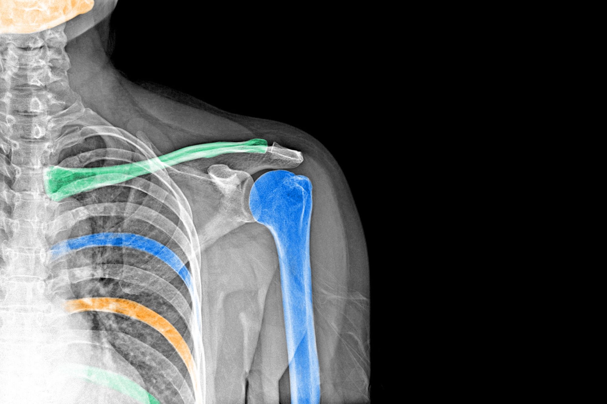
That high-energy attribute, while not exactly healthy for humans, is also what makes X-rays so useful. When X-rays hit an object, their energy is absorbed or scattered at different rates by different components of our bodies. X-rays have a harder time passing through bones, which show up as white on the resulting images, while they more easily penetrate our skin and internal organs, which show up darker.
X-rays revolutionized medicine upon their discovery, but that’s only one aspect of their amazing story. These six facts showcase just how important X-rays are to modern life, and how they’ve made the invisible visible.
The Discovery of X-Rays Was an Accident

Many of the world’s most important discoveries came about by accident, and you can add X-rays to that list. On November 8, 1895, German physicist Wilhelm Conrad Röntgen was experimenting with cathode rays in his laboratory in Würzburg when he noticed that a nearby screen had begun to glow. Not knowing what the mysterious rays causing the effect could be, he eventually called them “X-rays,” with the “X” referring to an unknown item (as in solving for “X” in mathematics). Röntgen noticed that these rays passed through soft tissue, like human skin, but didn’t penetrate harder materials such as metal and bone.
For the next seven weeks, Röntgen continued working in his lab in complete secrecy. When Röntgen’s wife asked what was the matter, he answered that if people knew what he was doing, they would say, “Röntgen must have gone mad.” Finally, on December 28 of that year, he published a paper titled “On a New Kind of Rays.” The world was never the same. Later, when asked what went through his mind when he first discovered X-rays, Röntgen answered: “I didn’t think; I investigated.”
The Discoverer of X-Rays Won the First Nobel Prize in Physics

In December 1896, a year after Röntgen published his groundbreaking paper, Alfred Nobel — famous for inventing dynamite — died in Sanremo, Italy, bequeathing his fortune to the establishment of a prize awarding the greatest advancements in literature, chemistry, physics, medicine, and peace (economics was added in 1969). Following five years of legal wrangling, in 1901 Wilhelm Röntgen became the first recipient of the Nobel Prize for physics, an award that eventually honored such titans of science as Albert Einstein, Marie Curie, and Niels Bohr. Röntgen also received an honorary degree of medicine from the University of Würzburg because of his invention’s immense medical benefits, but he never took out patents related to his invention.
People Used to Take “Bone Portraits” Using X-Rays

Many X-ray entrepreneurs and photo studios began offering “bone portraits” in the early years of the 20th century. The fad didn’t last, though, and that’s probably a good thing, because frequent, intense exposure to X-rays isn’t healthy for you. Those who regularly operated early X-ray machines developed skin lesions and other maladies because of prolonged exposure to ionizing radiation.
However, humans are constantly exposed to what’s called “background radiation.” The American Cancer Society estimates that today’s chest X-ray is the equivalent of 10 days of normal exposure — not terrible, but also not something you want to expose yourself to many times a day. So wearing those heavy X-ray vests probably isn’t a bad idea.
There Are “Hard” and “Soft” X-Rays

Not all X-rays are alike. Medical X-rays, CT scans, airport security scanners, and other devices most commonly associated with X-rays use what are known as “hard X-rays,” because they have smaller wavelengths and can therefore carry more energy. This makes them perfect for penetrating soft tissue to examine the harder structure lying underneath. Soft X-rays, on the other hand, have longer wavelengths, almost approaching the length of UV light. These X-rays can’t carry very much energy at all. However, these X-rays also have their uses in catalysis — the study of chemical reactions caused by catalysts — and biology.
X-Rays Helped Scientists Discover the Structure of DNA

On May 6, 1952, British chemist Rosalind Franklin at King’s College London took her 51st X-ray diffraction pattern of deoxyribonucleic acid, also known as DNA. For the first time in history, the image revealed DNA’s double helix structure. Known simply as Photo 51, the image had been produced by scattering X-rays off a pure fiber of DNA using a process known as X-ray crystallography.
Franklin’s colleague Maurice Wilkins showed the photo to two other scientists without her knowledge, and it was those three men who then won the Nobel Prize for medicine in 1962 — without any mention of Franklin’s contribution. (Sadly, she had passed away four years earlier.) Although she never received recognition from the Nobel Prize committee, scientists and historians now recognize her crucial contributions to molecular biology; the European Space Agency even named its Mars rover the Rosalind Franklin.
X-Rays Revolutionized the Study of Art History

Although X-rays have an obvious application in hospitals, art historians also have a need for technology that can delicately penetrate layers on a canvas to reveal the secrets beneath. X-rays are perfect at surpassing low-density materials to reveal high-density pigments (such as those containing metals like mercury, iron, zinc, and lead) below. This is particularly useful at uncovering underpaintings — the first layer of paint on a painting, often done historically with lead white — and other painted-over areas, which reveal an artist’s step-by-step approach to creating a masterpiece. The technology has been used to examine the works of Leonardo da Vinci, Vincent Van Gogh, and a variety of Dutch masters.